The debilitating effects of heat stress on poultry production have been well documented. Heat stress already results in severe economic loss worldwide. Regarding the decline in the reproductive performance of heat-stressed hens, the exact mechanisms involved are still unknown. This study was conducted to elucidate the molecular mechanisms underlying heat stress (HS)-induced abnormal egg-laying in laying hens. Hy-Line brown laying hens were exposed to HS at 32 °C or maintained at 22 °C (control) for 14 days. In addition, granulosa cells (GCs) from preovulatory follicles were subjected to normal (37 °C) or high (41 °C or 43 °C) temperatures in vitro. The results confirmed that laying hens reared under HS had impaired laying performance. HS inhibited proliferation, increased apoptosis, and altered the GC ultrastructure. HS also elevated progesterone secretion by increasing the expression of steroidogenic acute regulatory protein (StAR), cytochrome P450 family 11 subfamily A member 1 (CYP11A1), and 3b-hydroxysteroid dehydrogenase (3β-HSD). In addition, HS inhibited estrogen synthesis in GCs by decreasing the expression of the follicle-stimulating hormone receptor (FSHR) and cytochrome P450 family 19 subfamily A member 1 (CYP19A1). The upregulation of heat shock 70 kDa protein (HSP70) under HS was also observed. Collectively, laying hens exposed to high temperatures experienced damage to follicular GCs and steroidogenesis dysfunction, which reduced their laying performance. This study provides a molecular mechanism for the abnormal laying performance of hens subjected to HS, which may help when developing novel strategies to reverse the adverse impact.
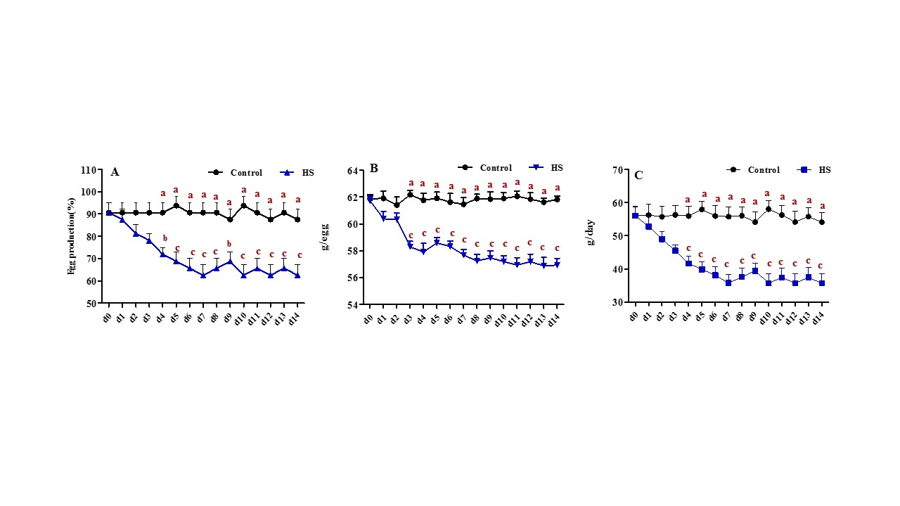
Figure 1. Effect of heat stress on the performance of laying hens. (A) Egg production, measured as percentages of laying hens/total hens. (B) Average egg weight of laying hens. (C) Egg mass, measured as g per egg produced/hen/D, on the day before heat stress (d0) and on each heat-stressed day (d1–d14) in control or stressed (HS) hens. The mean (± SEM) of 8 replicates, with each replicate containing 4 hens. Values with different letters are significantly different (p < 0.05).
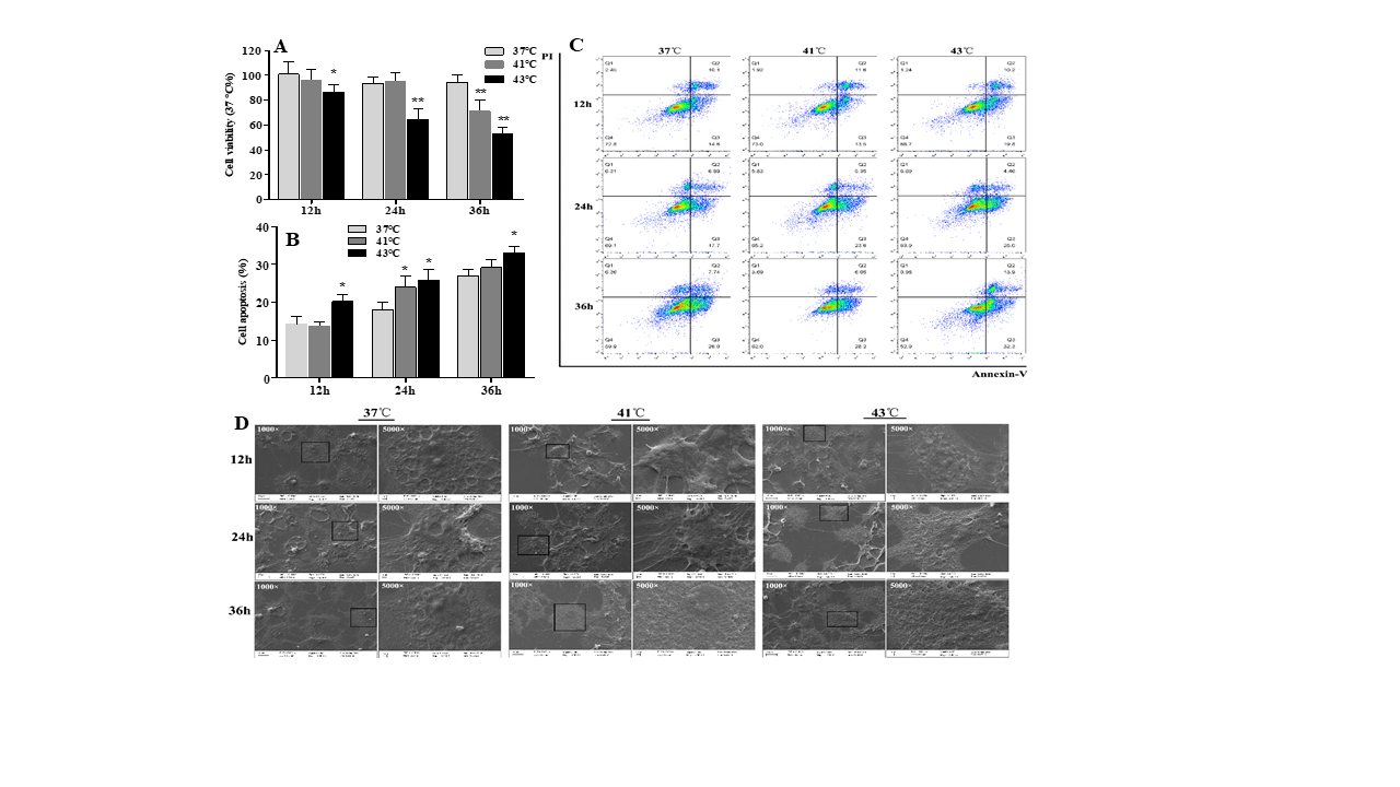
Figure 2. The effect of h eat stress on cell vitality and apoptosis of granulosa cells. Primary cultured chicken GC were kept at 41 °C or 43 °C for 12 h, 24 h, or 36 h. (A) CCK-8 assay. (B,C) Flow cytometric analysis, with cells in the area of the lower right corner (Q3) indicating FITC Annexin V-positive cells. (D) Scanning electron microscope after heat treatment for 12 h, 24 h, and 36 h. The boxed areas at 1000× were magnified to 5000× and were observed. * p < 0.05, ** p < 0.01.
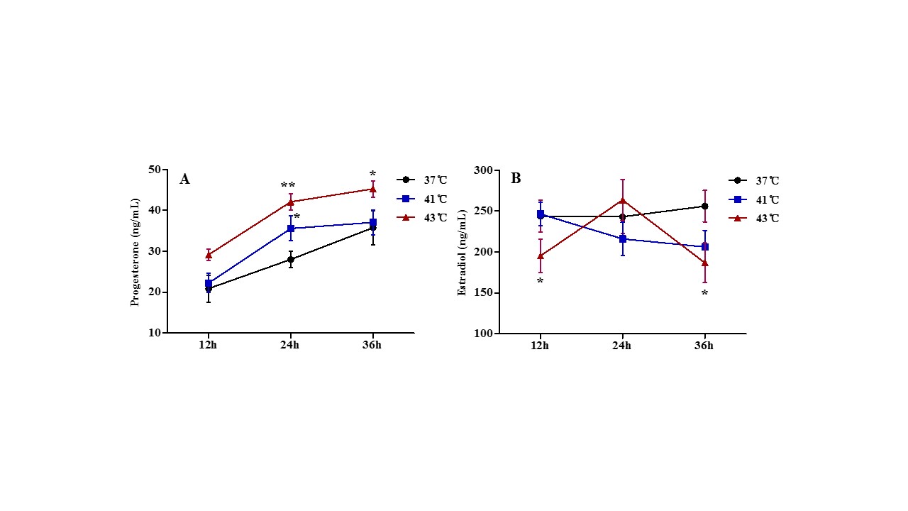
Figure 3. Effects of heat treatment on progesterone and estradiol synthesis in granulosa cells. Primary cultured GCs were kept at 37 °C, 41 °C and 43 °C for 12 h, 24 h, and 36 h, respectively. Progesterone (A) and estradiol (B) concentrations were analyzed using ELISA methods. * p < 0.05, ** p < 0.01.
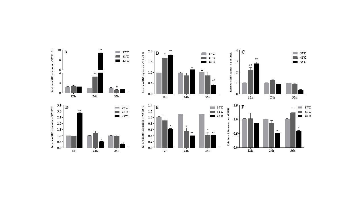
Figure 4. Effect of heat treatment on expression levels of progesterone and estradiol synthesizing enzymes in granulosa cells. (A–F) CYP11A1, StAR, 3β-HSD, CYP11A1, CYP17A1, CYP19A1, FSHR, CYP19A1, and FSHR mRNA expression levels, respectively. * p < 0.05, ** p < 0.01.
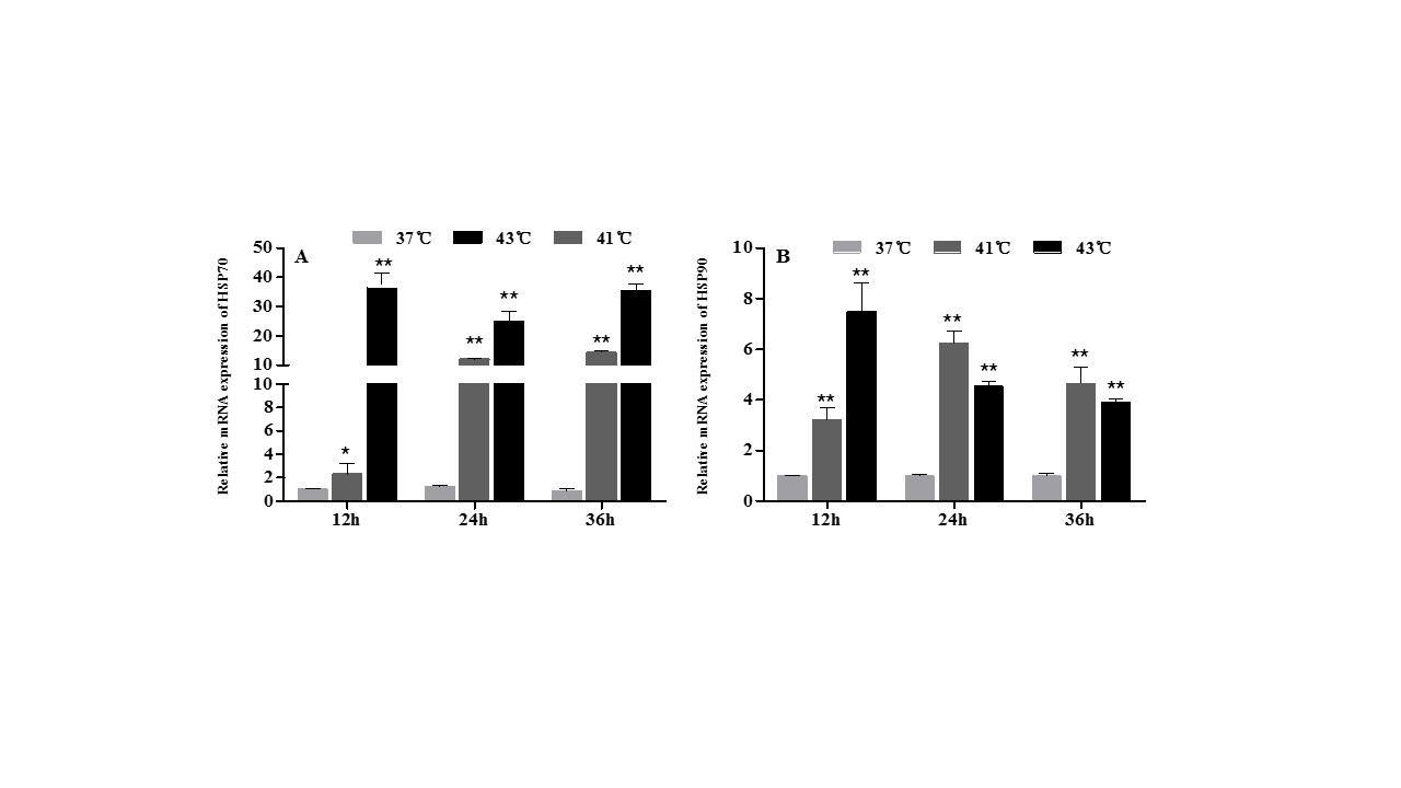
Figure 5. The effect of heat treatment on HSP70 (A) and HSP90 (B) mRNA expression levels in chicken granulosa cells. * p < 0.05, ** p < 0.01.
Read more:https://doi.org/10.3390/ani12111467


